
Colonoscopy to diagnose chronic ulcerative colitis in an 11-years-old Maltese
Abstract
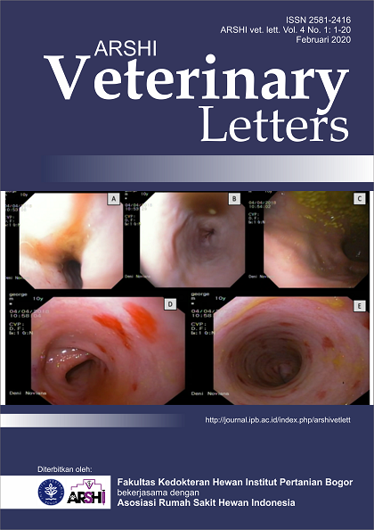
An 11-year-old castrated male Maltese was examined for increased frequency of defecation, mucus in feces, and chronic diarrhea with hematochezia. The dog was referred to Veterinary Teaching Hospital, Faculty of Veterinary Medicine, IPB University for further evaluation. Ultrasonography and colonoscopy were performed to further diagnose. Abdominal ultrasonography was taken using a linear probe with frequency 6-11 MHz. Colonoscopy was performed using colonoscope with tube length 700 mm and diameters 10 mm under anesthesia. Abdominal ultrasonography showed that the dog had a mucocele gall bladder, cholecystitis, hepatitis, slight-mild splenitis, nephrolithiasis, urolithiasis and thickened of the duodenal wall due to inflammatory bowel diseases. Colonoscopy showed ulceration and hemorrhage along the surface of the colon, whereas hyperemia only seen on the ascending colon. Based on endoscopic examination, the dog was diagnosed with severe and chronic ulcerative colitis. The authors recommended that the colonic biopsy should be undertaken in the dog presented with chronic ulcerative colitis.
Downloads
References
Argenta FF, de Souza SO, Meirelles LS, Snel GG, De Lorenzo C, Ienes-Lima J, Horn F, Driemeier D, Pavarini SP. 2018. Histiocytic Ulcerative Colitis in an American Staffordshire Terrier. Journal of Comparative Pathology. 165:40-44.
Cain CL, Bradley CW, Mauldin EA. 2017. Clinical and histologic features of acute-onset erythroderma in dogs with gastrointestinal disease: 18 cases (2005–2015). Journal of the American Veterinary Medical Association. 251(12):1439-49.
Cartwright JA, Breheny C, Major AC, Hill TL, Gow AG. 2016. Imaging diagnosis—a case of spontaneous hepatic portal vein gas in an 11‐month‐old west highland white terrier. Veterinary Radiology & Ultrasound. 57(5):E54-E57.
Davies DR, O'hara AJ, Irwin PJ, Guilford WG. 2004. Successful management of histiocytic ulcerative colitis with enrofloxacin in two Boxer dogs. Australian Veterinary Journal. 82(1‐2):58-61.
Stokes JE, Kruger JM, Mullaney T, Holan K, Schall W. 2001. Histiocytic ulcerative colitis in three non-boxer dogs. Journal of the American Animal Hospital Association. 37(5):461-465.
Copyright (c) 2020 CC-BY-SA

This work is licensed under a Creative Commons Attribution-ShareAlike 4.0 International License.
Authors who publish with this journal agree to the following terms:
1. Authors retain copyright and grant the journal right of first publication with the work simultaneously licensed under a Creative Commons Attribution License that allows others to share the work with an acknowledgement of the work's authorship and initial publication in this journal.
2. Authors are able to enter into separate, additional contractual arrangements for the non-exclusive distribution of the journal's published version of the work (e.g., post it to an institutional repository or publish it in a book), with an acknowledgement of its initial publication in this journal.
3. Authors are permitted and encouraged to post their work online (e.g., in institutional repositories or on their website) prior to and during the submission process, as it can lead to productive exchanges, as well as earlier and greater citation of published work (See The Effect of Open Access).